
In the first part of this blog I focussed mainly on the role of fungi in the decomposition of dead wood and referred only briefly in passing to some of the other organisms that thrive in the resource and habitat that is created when a tree dies and its trunk falls to the ground. Here I’ll focus on slime moulds, and in part 3 of the blog I’ll feature some of the other life forms that can readily be seen by looking closely at dead wood, with examples primarily from the Findhorn Hinterland area.

Slime moulds have some superficial similarities to fungi, but are actually part of a completely different Kingdom of life, and are as distinct from fungi as plants and animals are. They really need a new and improved common name, as their existing one, consisting of two words that both have negative connotations for many people, does a great disservice to what is actually a group of fascinating and remarkable organisms!

Slime moulds do have a scientific name of course – myxomycetes – but that’s hardly any more user-friendly than their common name. It’s a pity, because they deserve to be better-known and appreciated by people. Because of their small size and the fact that they mostly occur close to, or on, the ground, they are relatively inconspicuous, and therefore often overlooked. Although some species have very bright colours when they are in their sporulating phase, that only lasts for a few days at the most, so to see them it’s usually necessary to be in the right place at the right time.

I usually see most slime moulds in the late summer and autumn, as that is when many species are in their sporulating or reproductive phase. Going out at that time of year I’m always on the look-out for small patches of bright colour on dead wood, and, as with so many things, it gets easier once I’ve got my eye in for them and am familiar with the habitat where they are most likely to appear.

Slime moulds have complex life cycles, existing for part of their lives as separate, individual, single-celled organisms. At a certain stage, triggered by factors that are not entirely understood, those coalesce to form a multi-cellular plasmodium, which can flow and move across its substrate, often in the form of a branching network as seen in this photograph. This enables the slime mould to search for a food source and to find an optimum spot for the sporocarps or fruiting bodies to form and then release their spores, so that it has the best chance of reproducing successfully.

The fruiting bodies, which take the form of small spherical shapes in many species, then emerge out of the plasmodium, sometimes on stalks. This transition, from plasmodium to sporocarps, can be seen in the photographs here. When it is complete only the fruiting bodies will be visible, as the plasmodium will have been completely transformed by then, having served its function in the slime mould life cycle.


Once the sporocarps of a slime mould have released their spores their ecological function is complete and they decay and decompose very quickly. If they are encountered at this stage it can be very difficult, or impossible, to identify them to the level of species – that’s the case with the sporocarps of an unidentified slime mould in this photograph.

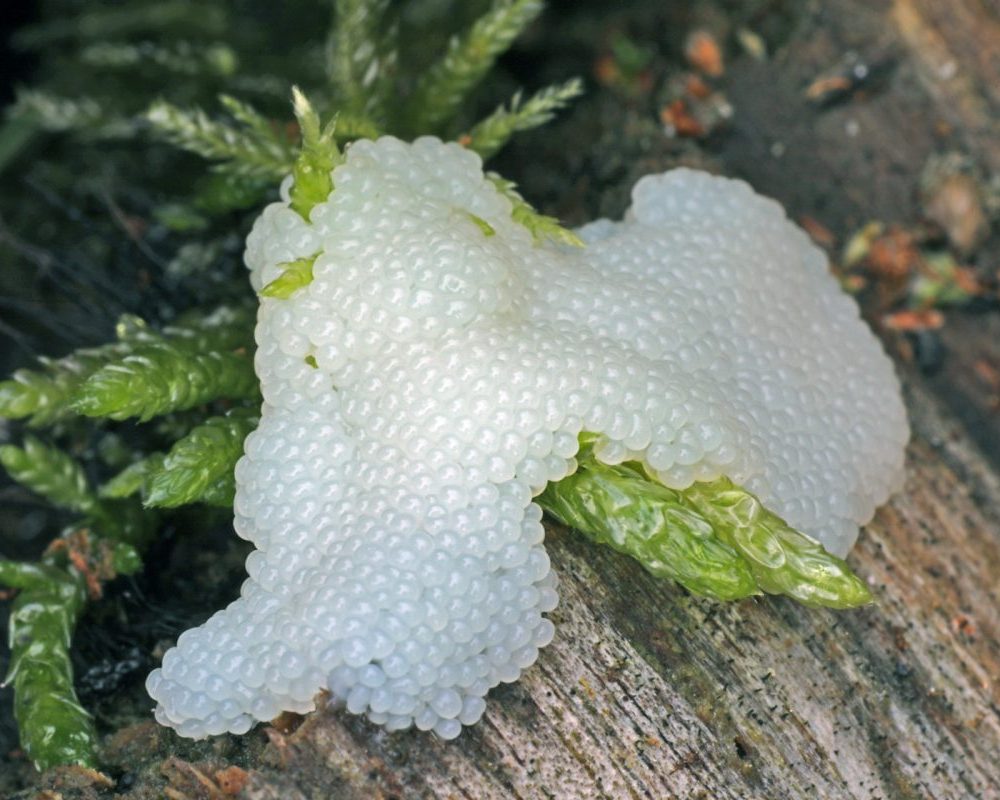
The sporocarps of some species are very different in their appearance, and a good example of this is provided by the slime mould Stemonitis fusca. When it enters its fruiting phase this species first becomes visible as a mass of whitish bubble-like spheres clustered together, as can be seen here.
Then, rather remarkably, out of these spheres emerge brown stalks bearing brown tubular structures called sporangia, within which the slime mould spores are produced. The two photographs below were taken 3 days apart, and show the transformation from the whitish spheres to the brown sporangia in Stemonitis fusca on a log in the Findhorn Hinterland.


After another day, the sporangia were fully developed, as shown in the photographs below. The left hand image shows the slime mould from the same angle as the previous photographs, whilst the photo on the right, taken from the other perspective, shows the structure of the sporangia more clearly.


These stalked sporangia are quite beautiful, especially when viewed close up using my high magnification macro lens:


Every patch of the slime mould has a unique cluster and arrangement of sporangia, making them as individual and varied from each other as each of us humans are different from one another. There’s an entire miniature world that displays the fantastic creativity of these remarkable organisms, but it’s highly ephemeral, as the fruiting bodies only exist for a very short period of time.
Sometimes the sporangia of this species fuse together, creating an amorphous, integrated brown mass, supported by the multiple stalks the sporangia have arisen from.


There is a wide and almost bewildering variety of forms that slime moulds manifest as, and here’s another couple of examples I’ve found on the Findhorn Hinterland:


Slime moulds complement the action of fungi in achieving the decomposition of dead wood and other plant material. Whereas fungi feed on and break down the lignin and cellulose of dead wood, slime moulds feed on micro-organisms such as bacteria and yeasts, and also on fungi themselves. These two entirely different groups of organisms can therefore often be seen close to each other on dead wood, with each one playing a specific and different role in helping to recycle the organic matter in the wood, transforming it into organic substances that can be used for the growth of other plants etc.
However, just as slime moulds will sometimes feed on fungal matter, so too there are fungi that feed on, and help to decompose, slime moulds, as these two examples from Trees for Life’s Dundreggan Conservation Estate in Glenmoriston illustrate:


In Part 3 of this blog, I’ll be featuring some of the mini-beasts that live in dead wood, including springtails, mites, millipedes and predators such as spiders, as well as various insects that overwinter as adults in old logs.
My thanks and appreciation to Bruce Ing for his expertise and assistance in identifying the slime moulds featured in this blog.
Leave a Reply